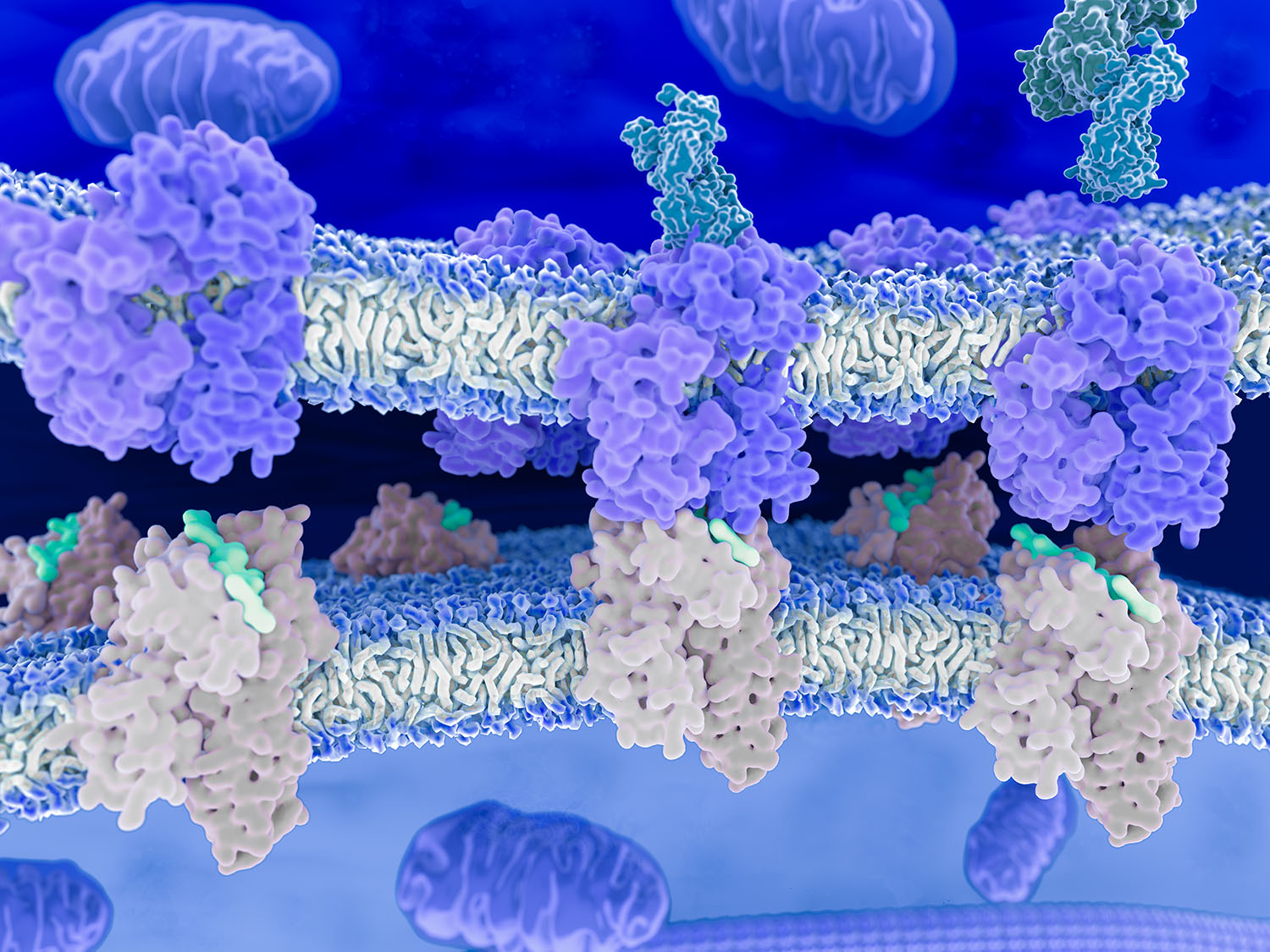
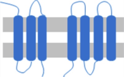
Membrane proteins
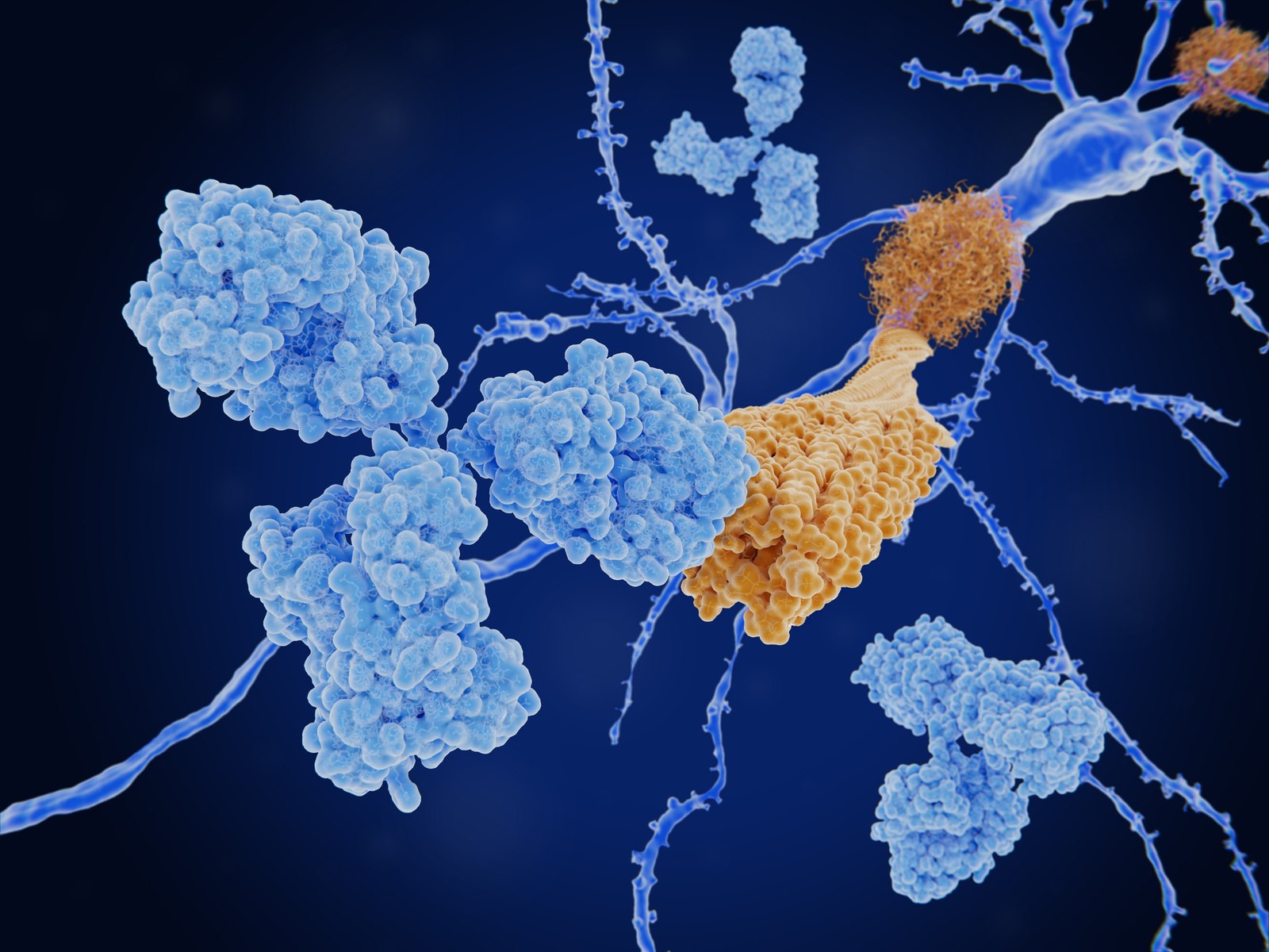
The leading challenge in therapeutic approaches against neurodegenerative diseases is the accurate characterization of the interaction between drug candidates and pathogenic oligomeric or fibrillar forms of proteins. MDS overcomes the limitations of surface-based methods which induce conformational heterogeneity and alter disease-relevant epitopes and provides KD values and stoichiometry necessary to understand the mechanism of action of drug candidates.
Overview
Neurodegenerative diseases (ND) affect millions of people worldwide and are characterized by the accumulation of neurotoxic amyloid aggregates in the brain leading to loss of function and ultimately death of neuronal cells. Immunotherapeutic approaches against NDs focus on antibodies that target the pathogenic aggregated forms of proteins such as α-synuclein or Aß peptide related to Parkinson’s disease and Alzheimer’s disease, respectively. ELISA and other surface-based methods are commonly used to identify antibodies that specifically target these pathogenic aggregates but tethering these sensitive proteins to a surface is often not feasible or induces artificial structures that do not occur in patients. Microfluidic diffusional sizing (MDS) avoids surface immobilization and thus preserves the disease-relevant structures of the aggregates which enables identification of antibodies that bind with high specificity and have the most effective mode of action against disease progression.
Purification-free quantification of affinity and receptor expression levels.
Case Study
Purification-free affinity and concentration measurement of membrane-protein targets
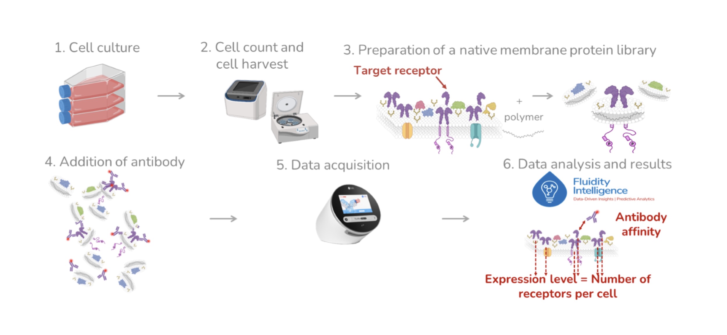
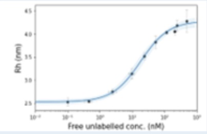
The improvement using MDS comes from the fact that we can produce this robust and precise data without much purification.

Lena Bauernhofer
Get started
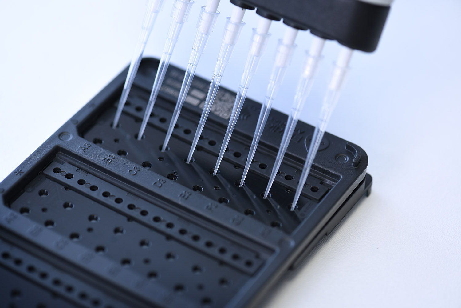
To study membrane proteins without purification, we recommend the following:
Workflow specification and benefits:
- 2 hours of sample preparation + overnight incubation (less than 30 minutes hands-on time)
- 2-3 hours for data collection and analysis
- KD range from pM to µM
- Concentration ranges from nM to µM
- Provides expression levels in terms of receptors per cell
- One standard T175 cell culture flask at 90–95% confluency will be sufficient for several experiments
- 12 µL of sample per triplicate
- Quick & easy to perform