Authors: Roland Worth1, Viola Denninger1, Sean Devenish1, Sebastian Fiedler1 and Tuomas Knowles1,2
1 – Fluidic Analytics, Cambridge, United Kingdom
2 – Centre of Misfolding Diseases, Yusuf Hamied Department of Chemistry, University of Cambridge, Cambridge, United Kingdom
Abstract
In this application note, we use microfluidic diffusional sizing (MDS) to measure size, affinity and stoichiometry of protein complexes formed by antibodies and intrinsically disordered proteins as well as their respective amyloid oligomers or fibrils. Our insolution approach allows for the direct use of disease-relevant proteins to understand antibody specificity and the mechanism of action. Specificity can be assessed by comparing the antibody affinity to neurotoxic, amyloidogenic aggregates vs. the monomeric (often non-toxic) species of the same protein. Stoichiometry is essential to determine the frequency of an epitope within protein aggregates which relates to a drug antibody’s mechanism of action. Moreover, our assay is very easy to perform, takes less than 3 hours in total, and requires only 4.0 μL per data point.

Introduction
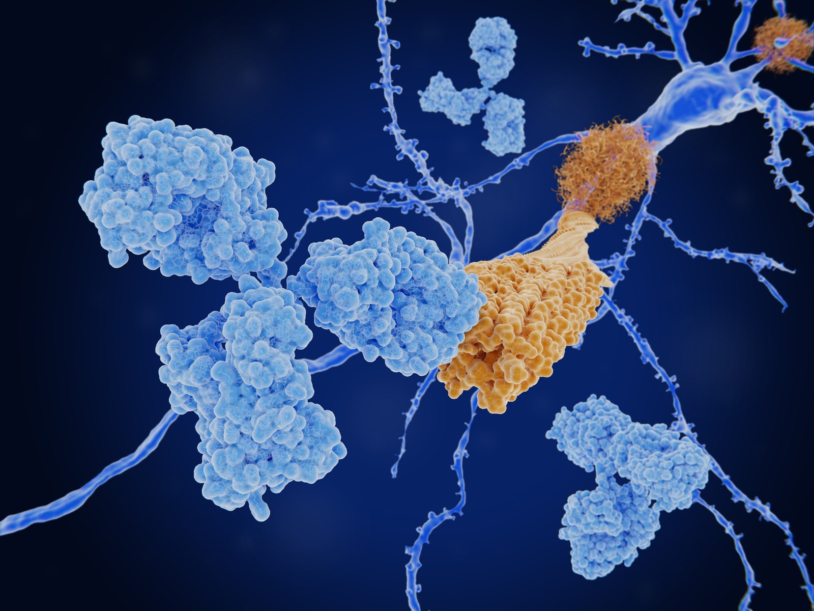
Formation of neurotoxic amyloid aggregates and their gradual accumulation in the brain are hallmarks of most neurodegenerative diseases (ND). While many efforts have been made to develop suitable therapeutics, to date there is no cure, and existing treatments only focus on reducing the symptoms and maintaining the quality of life of patients. Immunotherapy approaches against ND focus on antibodies that specifically target the pathogenic oligomeric or fibrillar forms of proteins like a-synuclein (Parkinson’s disease) or Aβ peptide (Alzheimer’s disease). Accurate characterization of the interaction between antibodies and these aggregate species, however, proves to be challenging for most technologies. For this reason, ELISA-based assays are still the method of choice to distinguish antibodies targeting conformational epitopes on the surface of oligomers or fibrils from those that bind to monomers. In a direct ELISA, monomers, oligomers, or fibrils are immobilized and tested for antibody binding (Figure 2). This approach, however, risks heterogeneous exposure of target epitopes due to surface immobilization.

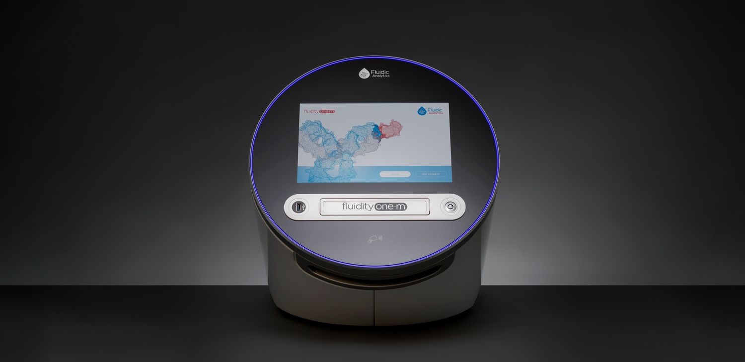
Another common ELISA assay to evaluate antibody specificity utilizes a competition format in which antibodies compete between surface-bound monomers and monomers, oligomers, and fibrils in solution. While this format measures the crucial interactions in solution and is thus less prone to surface-induced conformational heterogeneity it cannot provide universally comparable KD values or stoichiometry which are essential to elucidate the mechanism of action of a drug candidate. As shown in the case of aducanumab (Linse et al. 2020), a therapeutic antibody against Alzheimer’s disease, efficacy can be linked to stoichiometry of binding as revealed by MDS. Specifically, a more frequently exposed epitope causes oligomers or fibrils to be covered with antibody which impairs neurotoxicity of these amyloid aggregates. In this application note, we explain the principle of this new MDS assay including workflow, timings, and sample consumption using as an example a monoclonal antibody that binds to a-synuclein monomers, oligomers, and fibrils. Crucially, the assay only takes 2–3 hours, is immobilization-free, provides universally comparable KD values to assess specificity, and determines stoichiometry which is required to investigate the mechanism of action.
Results
To characterize the binding properties of 4D6 antibody to α-synuclein monomers, oligomers, and fibrils, it was first titrated against the monomeric form of α-synuclein. Using MDS, the hydrodynamic radius (Rh) of the free labeled α-synuclein monomer was measured to be 3.18 (± 0.12) nm (Figure 2). In the presence of antibody, the Rh was 5.01 (± 0.10) nm, therefore confirming complex formation. The measured sizes are typical of intrinsically disordered α-synuclein binding to a monoclonal antibody. Using our cloud-based Bayesian MAAP application, values were obtained for KD and stoichiometry (Figure 2). The results are presented as probability distributions with the mode (peak maximum) being the best estimate for KD and the width representing the uncertainty (error) associated with this value. The KD for monomer binding of 4D6 was determined to be 11.4 nM with a 95% highest density interval (HDI; similar to a 95% confidence interval) from 3.4 nM to 17.6 nM (Figure 3). In addition to KD, our assay simultaneously determines the binding stoichiometry in terms of how many monomers per antibody form the complex. In practice, this is achieved by simultaneously fitting the concentration of binding sites available for the fluorescently labeled species. The monomer stoichiometry is then calculated by the following equation:
stoichiometry mon = [binding sites]/[antibody]total.
In the case of 4D6 binding to monomer the stoichiometry is 2.2 (95% HDI 1.6–2.8) monomers per antibody as expected for antigen binding to a monoclonal IgG (Figure 3). The ability of 4D6 to bind to other forms of α-synuclein was determined by measuring equilibrium binding of fluorescently labeled 4D6 antibody and unlabeled α-synuclein oligomers or fibrils (Figure 2). In the presence of the α-synuclein oligomers or α-synuclein fibrils, the Rh of the complex was 7.86 (± 0.31) nm and 12.13 (± 0.38) nm, respectively. Thus, the antibody–fibril complex was considerably larger than the antibody–oligomer complex, confirming that Rh measurement by MDS is ideally suited to confirm the antibody bound to the species of interest. The affinities of 4D6 to oligomers and fibrils were very similar to those measured for monomer. While these binding experiments confirm that 4D6 binds various forms of α-synuclein with high affinity, our results suggest that the antibody likely binds to an epitope that is exposed at a similar level in the monomer, oligomers, and fibrils. We also determined binding stoichiometries of 4D6 to α-synuclein oligomers and fibrils. Unlike for the monomer experiments, binding to oligomers and fibrils was measured with fluorescently labeled antibody which is why the stoichiometry in terms of monomers per antibody is calculated as:
stoichiometryolig/fib = [ α-synuclein]total/[binding sites].
The stoichiometry values were 5.5 (95% HDI 3.7 – 8.2) and 7.1 (95% HDI 3.9 – 12.0) for oligomers and fibrils. These values can be interpreted as 1 antibody being bound every 5-6 α-synuclein monomers within an oligomer and 1 antibody being bound every 7 α-synuclein monomers within a fibril. These reduced binding stoichiometries relative to the monomer, suggest that within the oligomers and fibrils some monomers are unavailable for antibody binding, as would be expected.
Conclusion
Here we show that by using our novel in-solution assay based on MDS, we can accurately distinguish the binding targets for antibodies against amyloid aggregates such as a-synuclein monomer, oligomers, or fibrils. Crucially, the assay determines affinity and stoichiometry which are essential to evaluate antibody specificity and mechanism of action. Our in-solution assay takes less than 3 hours and uses low microgram amounts of protein.
Methods
Using a NanoDrop One™, the concentration of the a-synuclein monomer (ab218818, Abcam), oligomer (SPR-466B, 2BScientific) and fibrils (SPR-322B, 2BScientific) were determined in PBS (pH 7.4) by using the absorbance at 205 nm and an extinction coefficient of e205 = 31 mL.mg¯¹.cm¯¹, while the concentration of the 4D6 antibody (ab1903, Abcam) was determined in PBS (pH 7.4) at 280 nm using the IgG template (Thermo Fisher Scientific). For labeling, the a-synuclein monomer and 4D6 antibody were diluted into labeling buffer (0.2 M NaHCO3, pH 8.3) and mixed with Alexa Fluor™ 647 NHS ester (Thermo Fisher Scientific) at a dye-toprotein ratio of 3:1. Following incubation overnight at 4 °C, the labeled 4D6 antibody was purified using a 1 mL Pierce® Desalting Column (Thermo Fisher Scientific) with PBS (pH 7.4) as the elution buffer, while the labeled a-synuclein monomer was instead purified on an ÄKTA protein-chromatography system equipped with a Superdex™ 75 Increase 10/300 GL column (Cytiva, 29-1487-21) at a flow-rate of 0.5 mL.min-1 using PBS (pH 7.4) as a buffer. Prior to MDS measurement, a fresh batch of oligomers was thawed and used directly. Fibrils were sonicated prior to use to improve their homogeneity. Thawed fibrils were sonicated in a volume of 220 µL and a concentration of 0.56 mg.mL¯¹ using a Branson SFX150 Sonifier® (Fisher Scientific) using 20 s pulses for 6 min at an intensity of 80%. After sonication, the fibrils were centrifuged at 13000 x g for 20 min at 20 °C then filtered through a 0.22 µm Millex®-GV low protein binding Durapore® (PVDF) membrane filter. The concentration of the oligomers and fibrils were determined for each batch prior to use and in all cases refer to the concentration of the a-synuclein monomers making up the sample. For affinity measurements to the monomeric form of a-synuclein, the 4D6 antibody was diluted in PBS/0.05% Tween 20 (PBST) to give concentrations between 0.63–80 nM (2-fold dilutions). 4D6 was mixed with the fluorescently labeled a-synuclein monomer to give a final monomer concentration of 5 nM followed by incubation at 4 °C for 30 min. For affinity measurements to either the a-synuclein oligomers or fibrils, these were serially diluted in PBST to achieve concentrations of 3.5 nM– 3.54 µM (2-fold dilutions) or 1.2 nM–4.81 µM (2-fold dilutions), respectively. The labelled 4D6 antibody was then mixed with the a-synuclein samples to a final concentration of 5 nM and thereafter all samples were incubated for 30 min at 4 °C or room temperature for the a-synuclein oligomers or fibrils, respectively. Samples were measured at room temperature in triplicate on the Fluidity One-M using a size range of 3–14 nm for the a-synuclein monomers and oligomers, and 3.7–17 nm for the a-synuclein fibrils. The KD and antibody binding site concentration for all three interactions were determined by Bayesian inference (equations below).